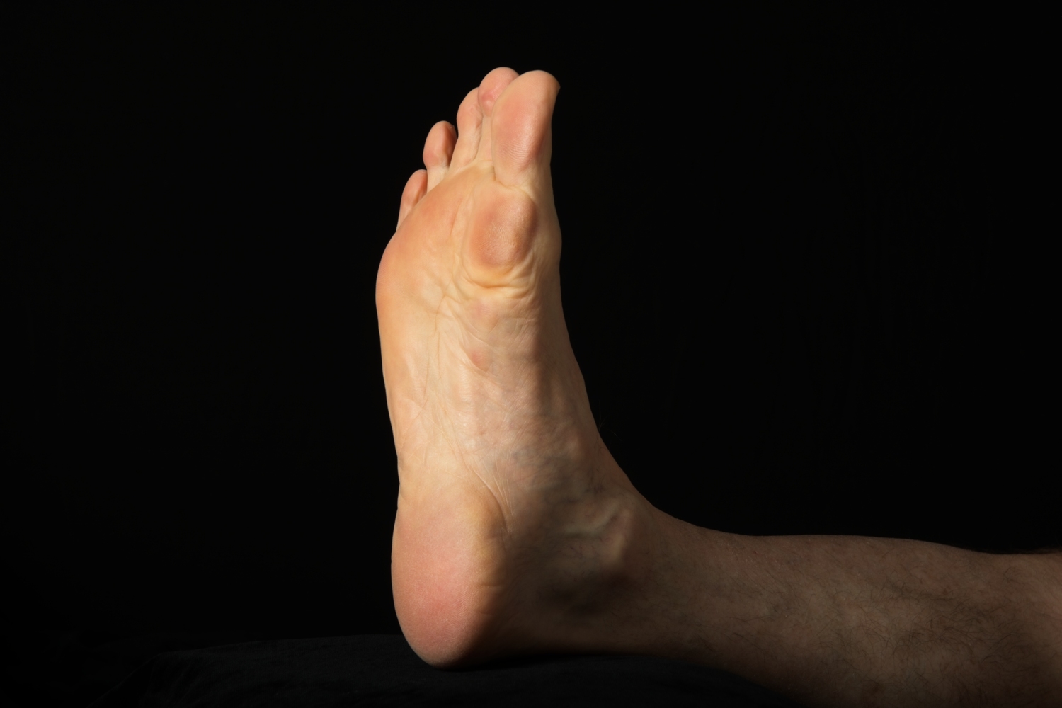

Harriet Thurlow is a podiatrist working within a busy Sydney private practice. She is endorsed to prescribe scheduled medicines and is a candidate to become a Credentialled Sports / Biomechanics podiatrist. Harriet sheds some light on plantar fibromatosis and its current treatment options.
As podiatrists, we are all too aware that plantar fibromatosis is difficult to diagnose and treat and can often present obscurely. Very broadly speaking, plantar fibromatosis is a benign fibrous tumour that arises from the plantar aponeurosis primarily the central and medial bands, which can be symptomatic or asymptomatic depending on the size of the lesion or lesions involved1,2.
Often referred to as ‘Ledderhose Disease’, this article will endeavour to shed some light on this condition and the current treatment options available.
Plantar fibromatosis is a condition which appears to affect primarily men who are middle aged2. It is related to, and presents concomitantly, with other hyperproliferative fibromatosis conditions such as Dupuytren’s Disease in the hands, Peyronie’s disease in the penis or people susceptible to keloid scarring. Interestingly, it has been associated with conditions such as frozen shoulder, excessive alcohol use, diabetes, epilepsy, smoking and repeated trauma3.
The nodules, or fibromas are isolated lesions which range in size from 0.5cm to 3cm in diameter and often present unilaterally3. Fortunately, they do not affect the surrounding musculature and skin. Thus, unlike the constriction of palmar fascia in Dupuytren’s Disease, contracture of the lesser digits and hallux is rarely seen3.
The development of nodules has been described to occur in three phases3. The first is a proliferative phase which is characterised by increased cellular proliferation and fibroblastic activity. Next is the active phase where increased collagen synthesis leads to nodular development, and finally, the residual phase where collagen maturation and scar formation occurs3.
For a large majority of patients, plantar fibromatosis initially presents as a painless condition where the primary symptom is a slow-growing lump along the inner portion of the arch3. In these early stages, symptoms may initially be described as local pressure and distension or a palpable but painless lump. However, as the fibromas increase in size, they can breach the critical level whereby load bearing activities become debilitating, and the area can become erythematic and painful4.
The space-occupying nature of plantar fibromas present significant mobility issues in those affected. Patients report some debilitation relating to barefoot activities, exacerbation of symptoms with restrictive shoes, or pain induced with any direct pressure on the fibroma3. These issues can be further intensified in the case of multiple fibromas developing over time3,4.
As with many other conditions, diagnostic certainty of plantar fibromatosis is important, as the possible differential diagnoses will have vastly different treatment plans and outcomes. In general, physical examination of the plantar fascia and hind foot will provide cues to either include or eliminate plantar fibromatosis.
The primary feature to identify a positive plantar fibromatosis diagnosis is the presence of single or multiple well-defined nodules along the whole length of plantar fascia. Concurrent reduced ankle dorsiflexion with tight posterior musculature may also be observed3. The possible differential diagnosis for plantar fibromatosis includes calcaneal stress fracture, tarsal tunnel syndrome, and plantar fasciitis3. These differentials can be distinguished by a thorough subjective history including description of pain, performing a calcaneal squeeze test, assessing for Tinel’s sign; as well as visually observing and palpating the plantar aspect of the feet, bony prominences and tendon insertions to eliminate other pain sources such as tendinopathies, bruising, skin breakdown or deformity3.
Typically, due to the obvious presentation of nodules in the plantar fascia, plantar fibromatosis is diagnosed within the clinical setting. However, if radiology is required, should the nodules be difficult to palpate or needing confirmation prior to commencing a treatment plan, then ultrasound or MRI are acceptable3. Ultrasound is the preferred modality due to time and cost effectiveness, but it can also provide an in-time comparison between both feet5. On MRI, the nodules appear as oval shaped areas of disorganisation originating from the plantar fascia3. The lesions are of low signal intensity in both T1 and T2 weighted sequences. Conversely, ultrasound allows sharp differentiation between the less reflective nodules and the more reflective plantar fascia5.
Due to the benign nature of plantar fibromatosis, many of the treatments are largely non-invasive. Surgical treatment is reserved for non-responsive, difficult to treat and painful lesions3. For podiatrists, the conservative treatment options may include offloading, using orthoses with apertures or cutouts to accommodate the nodules4. The aim for orthotic therapy is to reduce tension through the plantar fascia and avoid direct impingement of the fibroma4. Whilst this treatment is commonplace, there is limited evidence to support its efficacy. Further offloading may also be achieved by using rocker-bottom shoes to reduce the load of the foot, in gait, during midstance4. The goal for offloading through footwear and orthotics is to provide relief from the condition rather than influence the natural course of the disease3.
Extracorporeal shockwave therapy (ESWT) has been purported for use to soften the lesions by decreasing tissue contraction6. ESWT appears to be an effective option for management of plantar fibromatosis, even with treatment protocols being subject to variability6. Although further research is required, the current thinking is that ESWT stimulates biochemical signals like TGF-β and insulin-like growth factor 1 to be overexpressed6. This increased production of extracellular matrix components assists in counteracting the maturation process of myofibroblasts, resulting in less tissue contracture and ‘softer’ plantar fibromas, thus reducing pain6.
Corticosteroid injections are also demonstrating promising results for reducing inflammation and breaking down the fibrous tissue within the nodules. This form of treatment option is viable for an endorsed podiatrist to administer, or alternatively this treatment can be achieved with a referral to a radiologist. Corticosteroid injections are thought to decrease VCAM-1 expression which affects the production of cytokines TGF-β and FGF-β3. In a recent study by Flanagan et al. (2021), there were excellent outcomes with intralesional fenestration and Triamcinolone injections for treatment of plantar fibromatosis8. The current literature indicates that treatment should consist of between three to five intralesional injections using 15-30g of triamcinolone, four to six weeks apart. This of course is not without its risks, and skin and soft tissue atrophy and plantar fascia rupture need to be considered2.
Alternative treatments include radiotherapy, clostridium histolyticum collagenase, Verapamil, Tamoxifen. However, the complexities of these treatment options open discussions for another time, and currently lie outside a general podiatrist’s scope of practice3.
Surgical intervention is considered when the aforementioned treatments have not provided satisfactory relief. Whilst not in a general podiatrist’s scope of practice, there appears to be three main techniques used; local excision, wide excision and fasciectomies (either partial or complete)3. The recurrence rates reduce as the surgical technique used becomes more aggressive from 57-100% in local excision to 0-50% in complete fasciectomy, and as low as 6% in partial fasciotomies3. Of course, surgeries are not without their own complications and risk factors, which include impaired wound healing, painful scarring, skin necrosis and nerve entrapment9.
Plantar fibromatosis is a rare, benign condition where fibrotic nodules arise from the plantar fascia. Whilst often a painless condition, these nodules can increase up to 3cm in diameter. This condition is largely diagnosed clinically, but if radiology is needed, ultrasound is the recommended modality. There is growing evidence to support current treatment options, and clinicians can consider using orthoses, shockwave therapy, and cortisone injections in the preliminary treatment stages. Surgical options exist for aggressive, recalcitrant cases, or if painless function is not restored, although recurrence is not uncommon.
© Copyright 2021 The Australian Podiatry Association
